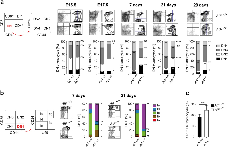
Fig. 5. Mitochondrial AIF loss led to the blockade of thymopoiesis at a DN immature state.
a Left, Gating strategy. Right, Cytometric identification of DN1-DN4 subsets of AIF+/Y and AIF−/Y embryos (E15.5 and E17.5) and 7-, 21-, and 28-day-old neonates (n = 7 embryos/mice per group). b Left, Gating strategy. Flow cytometry assessment of the DN1 prothymocyte compartment from 7- and 21-day-old AIF+/Y and AIF−/Y mice (n = 6 animals per group). c Frequency TCRβ chain positive DN thymocytes measured in AIF+/Y and AIF−/Y 21-day-old animals (n = 8 mice per group). Statistical significance was calculated by Mann–Whitney test. Bars represent mean ± SEM

