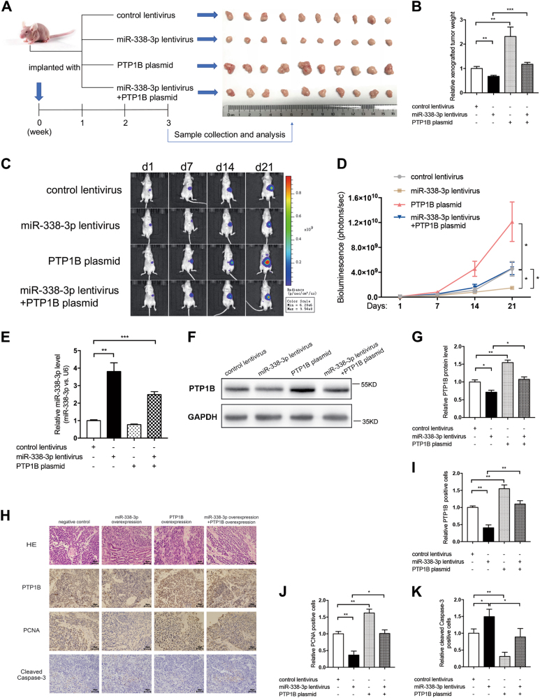
Fig. 7. miR-338-3p suppresses GC growth in vivo by targeting PTP1B.
a Flowchart of the experimental plan and representative images of xenograft tumors. MKN45 cells were infected with lentiviruses expressing firefly luciferase to construct MKN45-Luc cell line. MKN45-Luc cells were further infected with control lentivirus, miR-338-3p lentivirus, PTP1B plasmid, or miR-338-3p lentivirus plus PTP1B plasmid. Then, cells (1 × 10⁶ cells) were implanted into the subserosal area of the stomach of nude mice (10 mice per group) and the tumor growth was evaluated on day 21 after cell implantation. b Quantitative analysis of the tumor weight. c,d Bioluminescence imaging of tumors on day 1, 7, 14, and 21 after implantation of GC cells. c Representative images; d quantitative analysis. e qRT-PCR analysis of miR-338-3p levels in xenograft tumors. f,g Western blotting analysis of PTP1B protein levels in xenograft tumors. f Representative image; g quantitative analysis. h–k H&E and immunohistochemical staining for PTP1B, PCNA, and cleaved Caspase-3 in xenograft tumors. h Representative images; scale bar, 50 μm. i–k Quantitative analysis. n = 10 mice per group; data are represented as mean ± SEM; *p < 0.05; **p < 0.01; ***p < 0.001; two-tailed Student’s t-test

