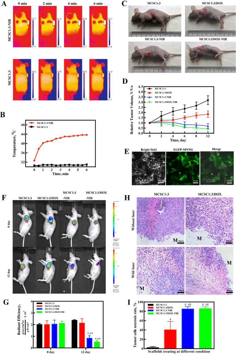
Figure 7.
The MCSC scaffolds exhibit robust anti-tumor effects in vivo. (A) IR thermal images and (B) temperature curve of MNNG xenograft mice exposed to NIR laser irradiation (red) or not (black). (C) Representative images for tumor volumes at day 12. (D) Relative tumor volume changes. (E) Lentivirus containing EGFP were successfully transfected in MNNG as indicated by fluorescence. (F) Representative images for the whole-body fluorescence imaging of tumors and (G) statistical analysis for intensity and area at day 0 and day 12. (H) Representative histology images of tumor tissues, M represents implanted scaffolds material. (I) Statistical analysis of tumor cell necrosis rate. *P < 0.05. IR, infrared.

