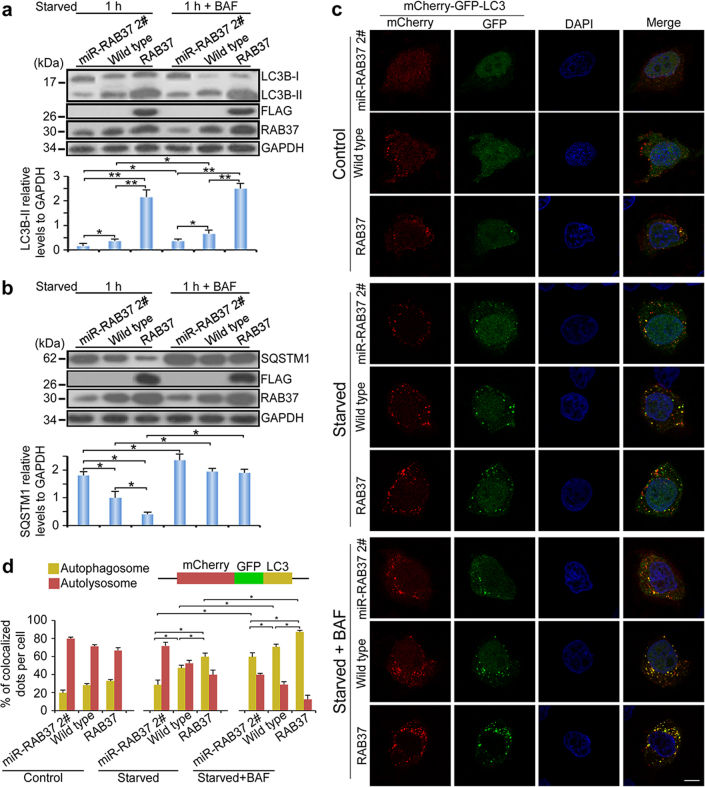
Fig. 7.
RAB37-associated autophagy flux. a, b RAB37 knockdown inhibits and over-expression promotes LC3B-II formation. Bafilomycin A1 treatment results in protein accumulation of LC3B-II and SQSTM1. RAB37 overexpression, RAB37 knockdown and control HeLa cells were cultured and then starved in EBSS with or without bafilomycin A1 (100 nM) for 1 h. The cell lysates were analysed by immunoblotting with antibodies as indicated. GAPDH was used as an endogenous control. Bottom: western blots were quantified for LC3B-II /GAPDH ratio and the SQSTM1/GAPDH ratio. Data are presented as means ± S.D. * stands for P < 0.05, ** stands for P < 0.01 (n = 3 independent experiments). c Autophagy flux revealed by the mCherry-GFP-LC3 tandem reporter in RAB37 knockdown (miR-RAB37 2#), over-expressed (stably expressing RAB37) and wild-type HeLa cell lines. Representative images of the cells transfected with the mCherry-GFP-LC3 reporter and grown in normal (control), EBSS medium (1 h) or EBSS with bafilomycin A1 (100 nM) addition (1 h) respectively. Single channel (red, green or blue) and merged images were taken by confocal microscopy. d Statistic analysis of vesicles positive for both GFP and mCherry (autophagosomes) and for mCherry (autolysosomes) (>15 cells per experiment) in (c). Percentages of co-localized dots (mCherry-GFP-LC3 (yellow) / mCherry -LC3 (red)) were counted. Data are presented as means ± S.D. * stands for P < 0.05, (n = 3 independent experiments)

