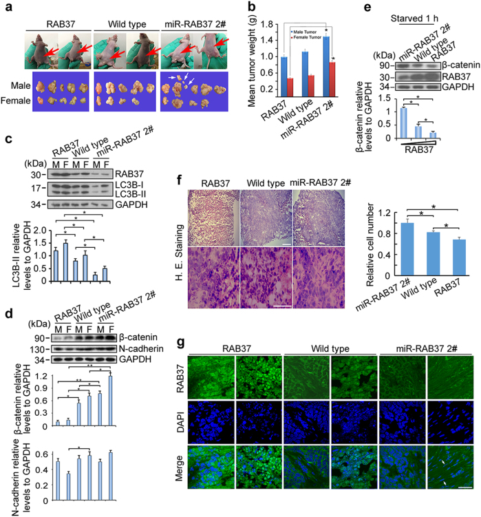
Fig. 8.
RAB37 promotes autophagy in vivo. a Nude mouse experiments. Three cell lines (HeLa cells stably expressing RAB37, miR-RAB37 2# and control cells) were used to implant nude mice. After 4 weeks, the tumours from both males and females were collected. Red arrows show tumour position in mouse. White arrows indicate tumour migration to other places in some mice. b RAB37 knockdown promotes tumour growth. Each bar represents the mean, and the error bars indicate the standard deviation. * stands for P < 0.05. c, d Tumour samples from three types of nude mice were examined through immunoblotting with anti-LC3B and anti-RAB37 (c), anti-β-catenin and N-cadherin antibodies (d). Both β-catenin and N-cadherin are EMT markers. GAPDH was used as an endogenous control. The molecular marker is showed on the left. Bottom: western blots were quantified for the LC3B-II/GAPDH, β-catenin/GAPDH, N-cadherin/GAPDH ratios. Data are presented as means ± S.D. * stands for P < 0.05, ** stands for P < 0.01 (n = 3 independent experiments). e RAB37 interference increases β-catenin protein level in HeLa cell culture in vitro. Bottom: western blots were quantified for β-catenin/GAPDH ratio. f Haematoxylin and eosin staining of three tumour types. Scale bar, 200 nm (up panels) or 100 nm (down panels). Relative cell numbers in one image was quantified. Data are presented as means ± S.D. * stands for P < 0.05, (n = 3 independent experiments). g Immunofluorescence analysis of tumour samples. Arrows show a longer tumour cell morphologically and a denser cell growth in RAB37-knockdown. The tumour sections were immunostained with an anti-RAB37 antibody (FITC). The nuclei were counterstained with DAPI (blue). The images were captured using confocal microscopy. Scale bar, 50 nm

