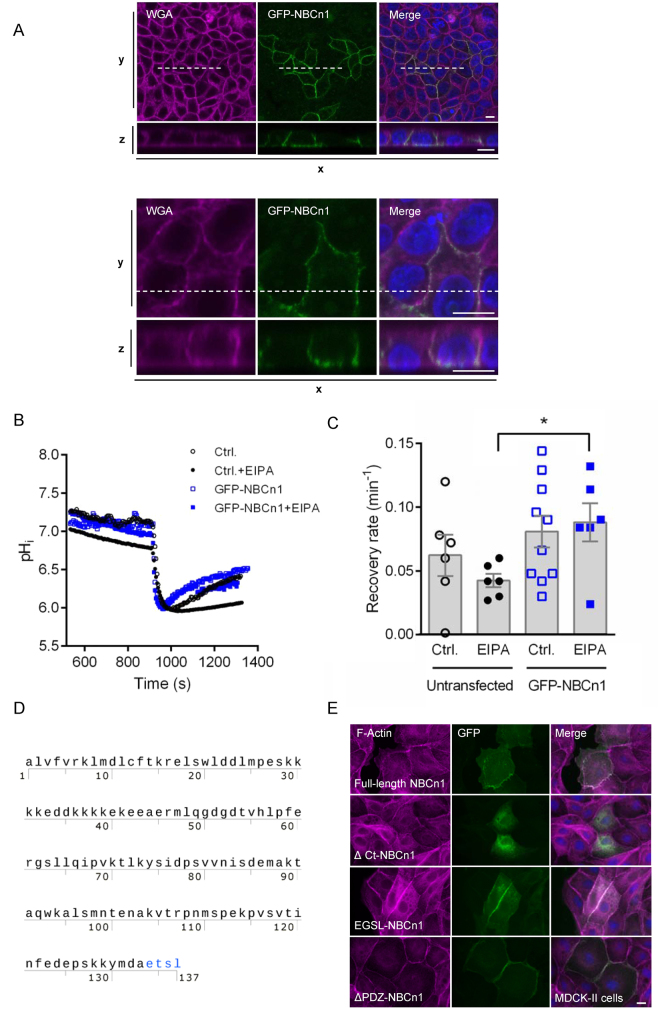
Figure 2.
GFP-NBCn1 membrane localization and function. (A) GFP-NBCn1 localizes to the basolateral cell surface. MDCK-II cells transiently transfected with GFP-NBCn1 were cultured to 100% confluency and maintained in culture for 12 days before fixation. Cells were stained with wheat germ agglutinin (WGA) and DAPI prior to confocal imaging. Bottom rows are xz-scans of the dashed line shown in top rows (xy-scans). Upper and lower panel are low- and high magnification images, respectively. Scale bars: 10 µm. Representative images from 4 independent experiments are shown. (B,C) GFP-NBCn1 is a functional net acid extruder. Capacity for net acid extrusion after an NH4Cl-prepulse-induced intracellular acidification was assessed by live imaging of BCECF fluorescence in GFP-NBCn1-transfected and control MCF-7 cells, in absence and presence of the NHE1 inhibitor EIPA. B, representative traces, C, Mean recovery rates. *p < 0.01, Student’s t-test, n = 6–10 per condition. (D) Amino acid sequence of the human NBCn1 C-terminal tail (Met1111-Leu1214), showing the PDZ-binding ETSL motif in red. (E) MDCK-II cells transiently transfected with GFP-coupled full-length NBCn1 (GFP-NCBn1), NBCn1 with deleted C-terminal (∆Ct-NBCn1), NBCn1 with a 4-amino acid deletion in the putative PDZ domain (ΔPDZ -NBCn1) or NBCn1 with mutated PDZ domain (EGSL-NBCn1). Fluorescence images of F-actin (magenta), GFP (green). Nuclei are stained with DAPI (blue). Scale bar 10 µm. Data are representative of 2–3 independent experiments per construct. Corresponding data for MCF-7 cells are shown in Fig. S2.

