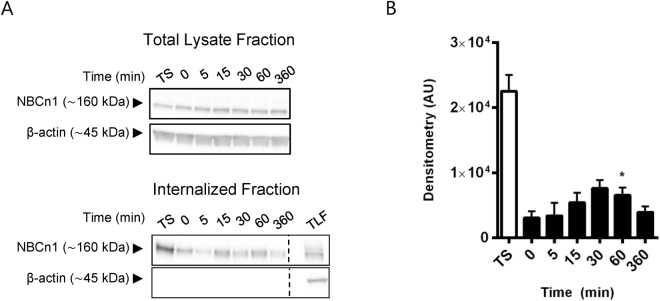
Figure 5.
NBCn1 is rapidly endocytosed from the basolateral membrane. MDCK-II cells were cultured on Transwell filters for 4 days and basolateral surface expressed proteins were labeled with cell impermeable biotin at 4 °C. Internalization was carried out at 37 °C, followed by stripping of biotinylated proteins still exposed at the cell surface. Cells were lysed and analyzed by Western blotting at the times indicated. (A) Representative Western blots of the total lysate fraction (top) and internalized (bottom) NBCn1. ß-actin was used as a loading control. TS: Total surface expression, TLF: total lysate fraction. For the 0 min time point, stripping off surface biotin was performed immediately after biotinylation. (B) Internalized NBCn1. Quantification of C showing mean raw densitometry (+SEM) of three independent experiments in arbitrary units (AU). *p < 0.05 One-way ANOVA with Dunnett’s post-hoc test (0 min is control column).

