Figure 6.
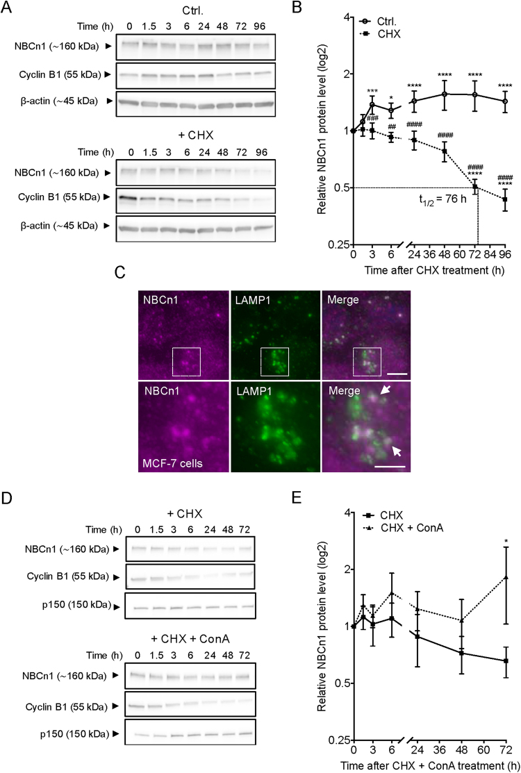
Protein turnover of NBCn1 is very slow. MCF-7 cells were cultured for 48 h prior to exposure to 100 µg/ml cycloheximide (CHX)+/− 10 nM concanamycin A (ConA) for the indicated time. Cells were lysed and processed for Western blotting (A,B and D,E) or fixed for immunofluorescence analysis (C). (A,D) Representative Western blots. ß-actin was used as a loading control and Cyclin B1 was used as a positive control for protein synthesis inhibition. (B,E) Protein turnover rate for NBCn1 after exposure to CHX+/− ConA. Values are normalized to 0 h for each exposure. (C) Fluorescence images of NBCn1 (magenta) and LAMP1 (green). Nuclei are stained with DAPI (blue). Scale bar 5 µm. Data are representative of 3–5 independent experiments. Western blot data are shown as mean with SEM error bars. *: comparison of values under the same treatment; #: comparison of values at the same condition but subjected to different exposures. */#, **/##, ***/### and ****/#### indicate P < 0.05, P < 0.01, P < 0.001 and P < 0.0001 respectively, relative to the initial value for that group (stars) or to the corresponding non-CHX-treated controls at the same time point (hash-tags). Two-way ANOVA with Bonferroni post-test. Similar data were obtained in MDCK-II cells (Fig. S5).

