Figure 1.
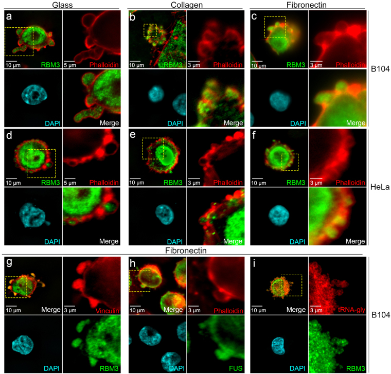
Localization of RBM3 to SICs in different cell types and plating conditions. Images of RBM3 (green), F-actin (red, phalloidin), and DAPI-stained nuclei (blue) in B104 cells (a–c) and HeLa cells (d–f) 30 minutes after plating onto glass, collagen, and fibronectin. For each cell type and substrate in panels a through f, upper right sub-panels show close-ups of F-actin distribution in regions (hatched yellow rectangles) at cell margins that contain SICs; sub-panels in lower right show the overlay of RBM3 with F-actin. (g–i) Images of B104 cells grown on fibronectin that were labeled for RBM3 and other SIC components by immunofluorescence, and for tRNA by FISH: cells were double labeled for (g) RBM3 (green) and the SIC component vinculin (red); (h) FUS (green) and F-actin (red); and (i) RBM3 (green) and tRNA-glycine (red).

