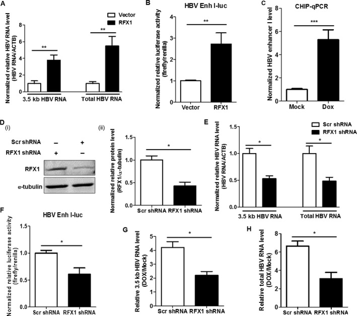
Figure 3.

RFX1 promotes HBV replication by enhancing the activity of HBV enhancer I. (A) HepG2 cells were cotransfected with pBB4.5‐1.2 × HBV expression plasmid (1 μg) and pCMV‐HA‐RFX1 expression plasmid or vector control pCMV‐HA (1 μg) using lipofectamine 2000 in a 35‐mm dish. The relative levels of 3.5 kb HBV RNA and total HBV RNA in the cells were quantified by qRT‐PCR. ACTB mRNA was used as an internal control. (B) HepG2 cells were cotransfected with pGL3‐HBV Enh I‐luciferase (Firefly), TRL‐PK (Renilla), and pCMV‐HA‐RFX1 expression plasmids or vector control pCMV‐HA using lipofectamine 2000 in a 12‐well plate. The effect of RFX1 in the activity of HBV enhancer I was detected by a dual‐luciferase assay. (C) HepAD38 cells were treated with 1 μmol/L Dox for 1 h, and the effect of Dox in the binding activity of RFX1 to HBV enhancer I (Enh I) was analyzed by ChIP‐PCR at 24 h post‐Dox treatment. (D) The levels of RFX1 protein in the pooled HepG2 cells stably transfected with RFX1 shRNA or scramble shRNA expression plasmid were detected by Western blot (i). The statistical graph (ii) of RFX1 protein levels detected by Western blot was analyzed by Image J software (NIH) at least in triplicate. The α‐tubulin was used as an internal control. (E) HepG2‐Scr and HepG2‐RFX1 shRNA cells were transfected with a pBB4.5‐1.2 × HBV expression plasmid (2 μg) using lipofectamine 2000 in a 35‐mm dish. The levels of 3.5 kb HBV RNA, total HBV RNA, and ACTB mRNA in cells were quantified by qRT‐PCR. ACTB mRNA was used as an internal control. (F) HepG2‐Scr and HepG2‐RFX1 shRNA cells were cotransfected with pGL3‐HBV Enh I‐luciferase and TRL‐PK using lipofectamine 2000 in a 12‐well plate. The effect of RFX1 in the activity of HBV enhancer I was detected by a dual‐luciferase assay. HepG2‐Scr and HepG2‐RFX1 shRNA cells were transfected with a pBB4.5‐1.2 × HBV expression plasmid (2 μg) using lipofectamine 2000 in a 35‐mm dish. After 24 h of transfection, the cells were treated with 1 μmol/L Dox for 1 h. The relative levels of 3.5 kb HBV RNA (G) and the total HBV RNA (H) in cells were quantified by qRT‐PCR at 48 h post‐Dox treatment. ACTB mRNA was used as an internal control.
