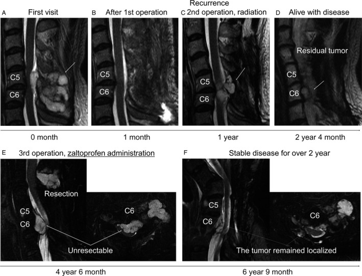
Figure 5.

(A, B) Sagittal, MRI T2 weighted image of cervical spine at first visit on the initial hospital before (A) and after (B) the surgery. The tumor (arrow) was resected (1st operation), which markedly compressed the spinal cord. (C) Local recurrence was detected at C5‐6 and tumor excision (2nd operation) was then performed after 1 year from the initial diagnosis. An arrow indicates the tumor. (D) The residual tumor (arrow). (E) Sagittal, MRI T2 fat suppression image. The tumors markedly enlarged and severe pain occurred in his left hand. The tumor of C2‐4 was resected, while the tumor of C5‐6 was unresectable. Zaltoprofen (240 mg/day) was then administrated for the pain. (F) Before the final operation for severe cervical kyphosis, MRI T2 fat suppression image showed the localized tumor of C5‐C6 remained.
