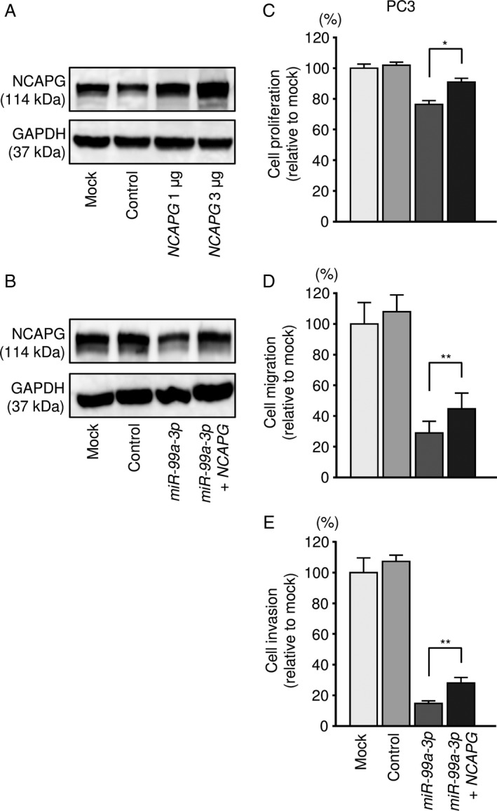
Figure 7.

Effects of cotransfection with NCAPG/miR‐99a‐3p in PCa cell lines. (A) NCAPG protein expression was evaluated by Western blot analysis of PC3 cells 48 h after forward transfection with the NCAPG vector. GAPDH was used as a loading control. (B) NCAPG protein expression was evaluated by Western blot analysis of PC3 cells 72 h after reverse transfection with miR‐99a‐3p and 48 h after forward transfection with the NCAPG vector. (C) Cell proliferation was determined using XTT assays 72 h after reverse transfection with miR‐99a‐3p and 48 h after forward transfection with the NCAPG vector. *P < 0.0001. (D) Cell migration activity was assessed by wound‐healing assays 48 h after reverse transfection with miR‐99a‐3p and 24 h after forward transfection with the NCAPG vector. **P < 0.001. (E) Cell invasion activity was characterized by invasion assays 48 h after reverse transfection with miR‐99a‐3p and 24 h after forward transfection with the NCAPG vector. **P < 0.001.
