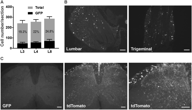
Figure 1.
Distribution of GFP-positive cells in lumbar DRGs and spinal cord following intraplantar administration of AAV2/9-CBA-Cre-GFP at postnatal day 5. (A) Quantification of GFP positive cells in lumbar dorsal root ganglia (L3-5) following the intraplantar delivery of AAV2/9-CBA-Cre-GFP. Data are the mean +/− S.E.M. of 3 mice for which the % of GFP-labeled neurons observed in 8 to 18 sections per mice has been averaged. (B) Representative photomicrographs illustrating GFP distribution in lumbar and trigeminal ganglia. (C) Representative photomicrographs illustrating GFP (absence of) and tdTomato staining in the lumbar spinal cord of an animal co-injected with AAV2/9-CBA-Cre-GFP and emCBA-Flex-tdTomato-WPRE are shown. Scale bars = 100 µm.

