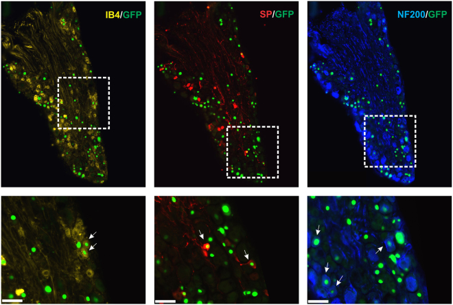
Figure 2.
Distribution of the Cre-GFP in the neuronal subpopulations within the lumbar dorsal root ganglia. Representative photomicrographs showing co-localization of GFP with markers of peptidergic (Substance P), non-peptidergic (Isolectine B4) and large diameter myelinated (NF200) neurons are shown for mice (n = 6) injected in the plantar surface of both hindpaws with the AAV2/9-CBA-Cre-GFP virus. Arrows indicate neurons co-labeled for GFP and the indicated marker. Scale bars = 50 µm.

