Figure 4.
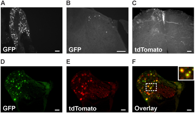
Distribution of GFP-positive cells in lumbar dorsal root ganglia and spinal cord following the intrathecal administration of AAV2/9-CBA-Cre-GFP. Representative photomicrographs illustrating GFP distribution in lumbar dorsal root ganglia (A) and the lumbar spinal cord (B, the dorsal horn is shown) of a mouse injected intrathecally with the AAV2/9-CBA-Cre-GFP virus at the adult age (25 days old; n = 4 mice). A representative photomicrograph illustrating the expression of tdTomato in the lumbar spinal cord of an animal co-injected with AAV2/9-CBA-Cre-GFP and AAV-emCBA-Flex-tdTomato-WPRE is also shown (C). (D–F) Nuclear GFP staining and tdTomato expression in a lumbar DRG of an animal co-injected with AAV2/9-CBA-Cre-GFP and AAV-emCBA-Flex-tdTomato-WPRE are shown. For panels C–F, images are representative of what has been observed in 3 mice. Scale bars = 100 µm.

