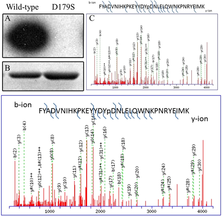
Figure 2.
Autophosphorylation activity of PfCK2. (A) Autoradiograph kinase activity assay was carried out using 2 μg of wild type (lane 1) or D179S mutant PfCK2 (lane 2) with Mg [γ32P]-ATP. (B) The proteins were resolved by SDS-PAGE, stained with Coomassie blue and their activity analysed by autoradiography. (C) Annotated MS/MS spectra showing identification of Y30 phosphorylation using the Mascot protein database search software. The Tyr30 (labeled Y16 in the spectrum) is unambiguously identified as it is sandwiched by a series of y-ions [y16,y17,y18(phosphorylation site),y19,y20]. y13 ion was trimmed 10 times to show the weaker ions in the spectrum.

