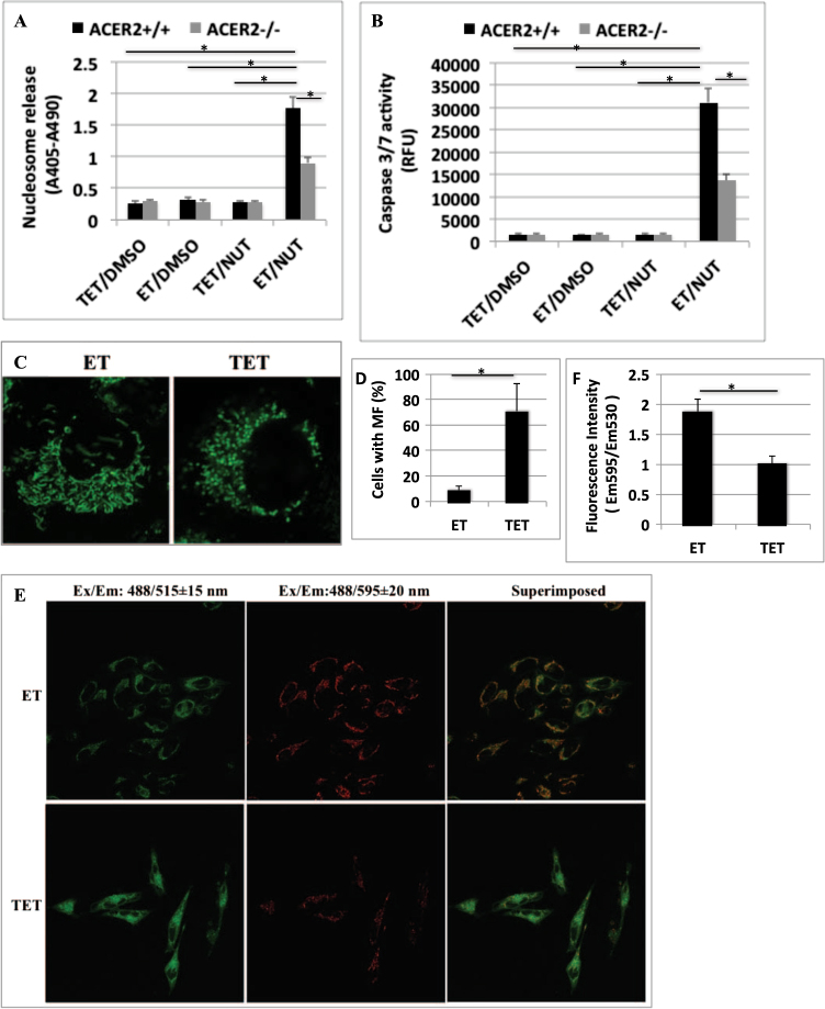
Fig. 4. High ACER2 upregulation mediates PCD in response to hyperactivation of p53.
a, b H1299:p53TRE/ACER2+/+ cells or H1299:p53TRE/ACER2−/− cells were grown in the presence of ET plus DMSO, ET plus NUT (10 μM), TET (1 μM) plus DMSO, or TET plus NUT for 72 h before DNA fragmentation a and caspase 3/7 activity b were determined. c, d HeLa:ACER2TO cells were grown in the presence of ET or TET (100 ng/ml) for 48 h before mitochondria were imaged under the confocal microscope C and the percentages of cells with MF were computed d. e, f HeLa:ACER2TO cells were grown in the presence of ET or TET (100 ng/ml) for 48 h before being subjected to MMP assays by confocal microscopy e or fluorometry f as described in Methods section. The data represent mean values ± SD of three independent experiments. Statistical analyzes were performed as described in Methods section. *represents p < 0.001

