Figure 1.

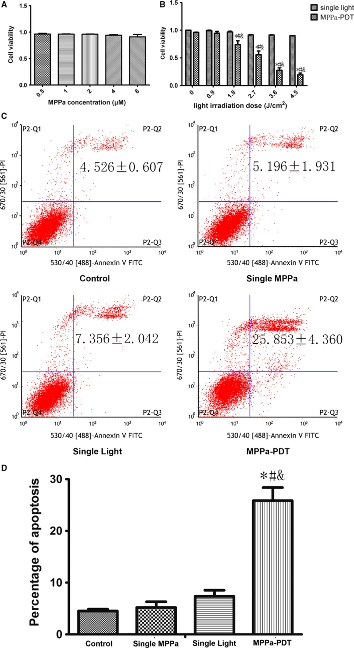
MPPa‐PDT produced cytotoxicity and apoptosis in MDA‐MB‐231 cells. (A) Cell viability of MPPa at different concentrations detected by the CCK‐8 assay. (A) Cell viability of MPPa at 2 μM and different does of light detected by the CCK‐8 assay. (C) Apoptosis detected by annexin V/PI double‐staining. (D) Percentage of apoptosis. *P < 0.05, MPPa‐PDT vs. control. #P < 0.05, MPPa‐PDT vs. Single MPPa; &P < 0.05 MPPa‐PDT vs. Single light. Values are mean ± SD of three independent determinations.
