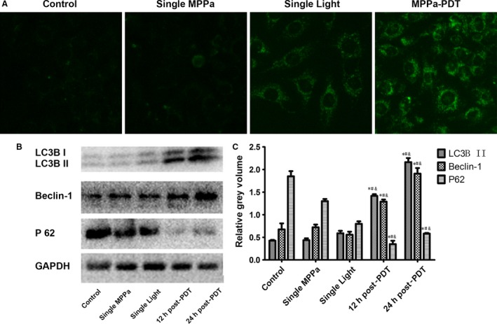
Figure 2.

MPPa‐PDT induces autophagy. (A) The expression of autophagic vacuoles detected by immunofluorescence was observed by monodansylcadaverine (MDC) staining (MDC,×200). MDC (green) staining. (B) Expression of LC3B II, Beclin‐1, P 62 proteins Lanes; (C) Relative Gray volume. *P < 0.05, MPPa‐PDT vs. control. #P < 0.05, MPPa‐PDT vs. Single MPPa; &P < 0.05 MPPa‐PDT vs. Single light. Values are mean ± SD of three independent determinations.
