Figure 6.
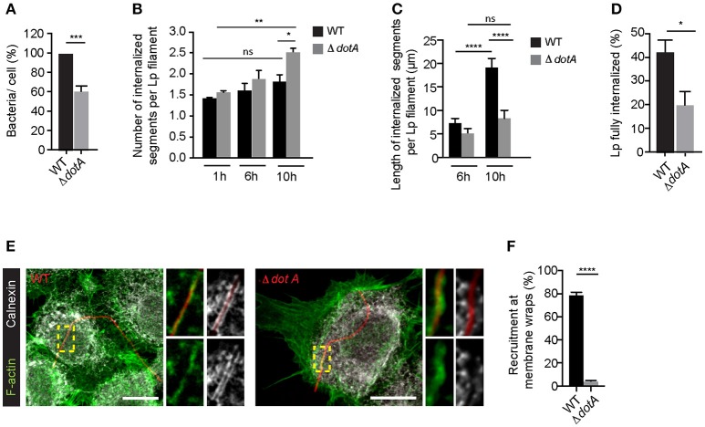
Dot/Icm translocated effectors are required for the attachment and entry of filamentous Lp in LECs (A) Quantification of the efficiency of attachment of dotA mutants compared to wild-type Lp 1 h p.i. Data shown are mean ± SEM from 5 independent experiments, normalized to wild-type controls (n > 500 cells per experiment). Statistical analysis was performed using Student's. ***p < 0.001. (B) Number of internal segments for wild-type and dotA mutants. Cells infected with RFP-WT or RFP-dotA mutants were washed at 1 h p.i to remove unbound bacteria and fixed or internalization was allowed to proceed for additional 5 or 9 h followed by immunolabelling of external bacteria. Each segment undergoing internalization was considered as a separate event. Data shown are mean ± SEM from 3 independent experiments (n > 100 in each experiment). Statistical tests were performed using one way- ANOVA with Tukey's multiple comparison test, *p < 0.05. (C) Internalization of dotA mutants compared to wild-type Lp 6 and 10 h p.i. Lengths of bacteria internalized were measured using differential immunolabeling to label external bacteria. Data shown are mean ± SEMs from 3 independent experiments (n > 100 bacteria in each experiment). Statistical tests were performed using one way- ANOVA with Tukey's multiple comparisons test, ****p < 0.0001. (D) Efficiency of bacterial internalization at 12 h p.i. Cells infected with RFP-WT or RFP-dotA mutants were washed 1 h p.i. to remove unbound bacteria. Cells were fixed 12 h p.i and external bacteria were immunolabeled. The number of bacteria fully internalized was quantified in three individual experiments. Data shown are mean ± SEM from 3 independent experiments (n>100 in each experiment). Statistical analysis was performed using Student's, *p < 0.01. (E) Representative confocal micrographs showing the recruitment of calnexin (grayscale) and F-actin (green) at the membrane wraps formed by attachment of RFP-WT (left) or RFP-dotA mutants (right) 6 h p.i. For all fluorescence micrographs the main panels are merged z-stacks and the higher magnifications are single z-planes. All scale bars, 12 μm. (F) Quantification from (E). Data shown are means± SEMs from 3 independent experiments (n > 25 in each case). Statistical analysis was performed using Student's t-test, ****p < 0.0001.

