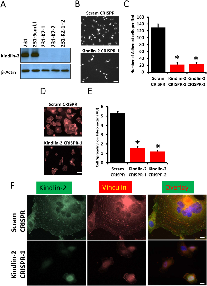
Figure 1.
Kindlin-2 is required for cancer cell adhesion, spreading and focal adhesion formation. (A) Western blots developed with anti-Kindlin-2 antibody of protein lysates from MDA-MB-231 transduced with a scrambled sgRNA (Scram CRISPR), Kindlin-2 sgRNA-1 (K2 CRISPR-1), Kindlin-2 sgRNA-2- (K2 CRISPR-2) or both sgRNA-1 and -2 (K2-CRISPR-1 + 2). β-Actin is a loading control. (B,C) Cell adhesion assay: Control (Scram CRISPR) or Kindlin-2-deficient (Kindlin-2 CRISPR-1 and -2) MDA-MB-21 cells were seeded onto on fibronectin-coated slides for 1 h, at which point they were fixed and stained with phalloidin-568 to visualize actin filaments. Micrographs of K2-CRISPR-1 are shown as an example in A and quantification is shown in B. (D,E) Cell spreading assay: Control (Scram CRISPR) or Kindlin-2-deficient (Kindlin-2 CRISPR-1 and -2) MDA-MB-21 cells were seeded onto on fibronectin-coated slides for 2 h, at which point they were fixed and stained with phalloidin-568 to visualize actin filaments. Micrographs of K2-CRISPR-1 are shown as an example in D and quantification is shown in E. (F) Representative confocal microscopy micrographs of Control (Scram CRISPR, top panels) or Kindlin-2-deficient (Kindlin-2 CRISPR-1, bottom panels) MDA-MB-21 cells that were stained for Kindin-2 (left panels) and Vinculin (middle panels). Co-localization of both Kindlin-2 and Vinculin at the focal adhesion structures is shown as yellow puncta in the overlay right panels. Data are the means ± SEM, N = 3; *p < 0.05; Student’s t-test).

