Figure 7.
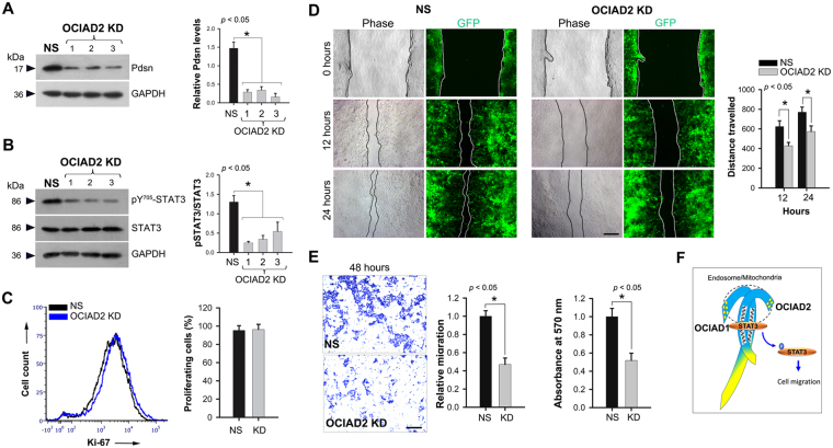
Knockdown of OCIAD2 retards migration of HEK293 cells. (A) Western Blotting confirming ociad2 knockdown (KD) in OCIAD2_ shRNA (1, 2 and 3) transfected HEK293 cells as compared to non-silencing (NS) shRNA. Graph shows relative OCIAD2 levels in NS and KD. n = 4 (B) Western Blotting for detection of STAT3 and pSTAT3 (Y705) levels in NS and KD. Data are representative of three independent experiments and graph shows relative STAT3/pSTAT3 ratio upon ociad2 KD. Protein size markers (in kDa) are indicated to the left. Full-length blots are presented in Supplementary Figure S6. n = 4 (C) Flow cytometry analysis of Ki-67+ population in ociad2 KD HEK293 cells. Graph shows results obtained from four independent experiments (mean ± SEM). (D) Reduced migration of ociad2 KD cells observed in wound healing assays. Monolayers of control and ociad2 knockdown HEK293 cells were wounded and the degree of recovery was measured at 0, 12 and 24 hours post-wounding. Representative phase and fluorescence images, 4× magnification. Measurement and estimation of wound recovery was based on the initial wound size. Graph shows quantification of results obtained from three independent experiments (mean ± SEM) with at least 5 fields analyzed per sample per experiment. (E) Representative images (4× magnification) showing reduced migration of ociad2 KD HEK293 cells after 48 hours in a trans-well migration assays. Graphs represent quantification of cell migration (absorbance at 570 nm) obtained from two independent trans-well experiments performed in two technical replicates (mean ± SEM). *p < 0.05 was used to indicate a statistically significant difference. (F) Proposed model depicting the location of OCIAD1 and OCIAD2 and their interaction via the double helical motif (wavy grey lines). Blue shading: conserved region; Yellow: non-conserved regions. STAT3 interaction with OCIAD1 or OCIAD2 (individually or together) leads to STAT3 activation essential for cell migration.

