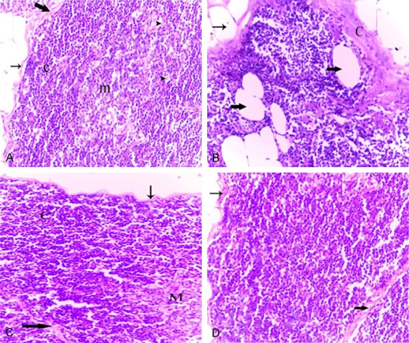
Figure 5.

Photomicrographs of thymus tissues of normal adult rat (A), normal aged (B), normal aged treated with either melatonin (C) or turmeric (D). (A) Thymus tissues of normal adult rats showing a dark stained cortex (c) with densely backed lymphocytes and light stained medulla (m) with lymphocytes and macrophages (short arrows) as well as epithelial cells (irregular arrows). Note the thin capsule (thin arrow) and the thin interlobular septum (thick arrow). (B) Thymus tissues of normal aged rats showing the lymphocytes depletion of the cortex. Note the thickened capsule (C) and increased numbers of adipocytes infiltered the capsular (thin arrow) and extended into the cortex (thick arrows). (C) Thymus of a normal aged rat treated with melatonin showing increased lymphocytic population in the cortical (C) and medullary (M) regions. Note the thin capsule (thin arrow) and interlobular septum (thick arrow), as well as the absence of adipocytes. (D) Thymus of a normal aged rat treated with turmeric showing the histological improvement in the thymic tissue. Note: the cortical (C) and medullary (M) regions. (H&E, 400X).
