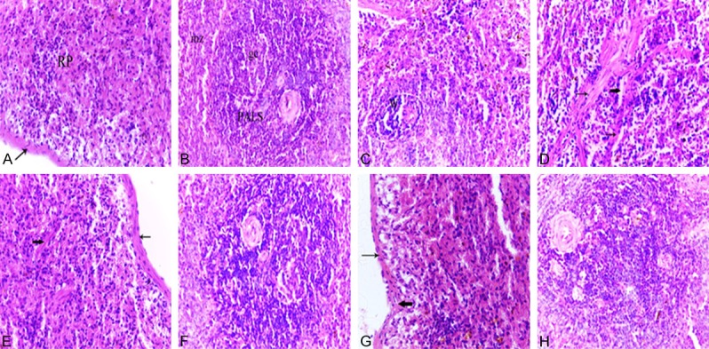
Figure 6.

Photomicrographs of spleen tissues of a normal adult rat (A, B), normal aged (C, D), normal aged treated with either melatonin (E, F) or turmeric (G, H). (A) Spleen of an adult rat showing the splenic red pulp (RP). Note the less-thickened capsule (arrow). (B) Spleen of an adult rat showing the PALS and marginal zones (mz). Note the presence of follicles containing prominent germinal centers (gc) with tangible body macrophages which appeared with cytoplasmic engulfed apoptotic debris. (C) Spleen of a normal aged rat revealed the decreased cellularity of both white pulp and red pulp areas. Note the lymphocytic depletion of the white pulp (W) and the marginal zone is hardly defined. (D) Spleen of a normal aged rat showing the lymphocytic depletion of the white pulp (W) with shrinkage and apoptotic cells. Note the tangible body macrophages with cytoplasmic engulfed apoptotic bodies. (H&E, 400X). (E) Spleen of a normal aged rat treated with melatonin showing the increased lymphocytic population of the splenic red pulp. Note the less-thickened capsule (thin arrow) and trabeculae (thick arrow). (F) Spleen of a normal aged rat treated with melatonin showing a marked improvement where the lymphocytic population of the white pulp was increased. (G) Spleen of a normal aged rat treated with turmeric showing the increased lymphocytic population of the splenic red pulp. Note the less-thickened capsule (thin arrow) and trabeculae (thick arrow) and the decrease number of pigments. (H) Spleen of a normal aged rat treated with turmeric showing apparent increase in the lymphocyte population of white pulp. (H&E, 400X).
