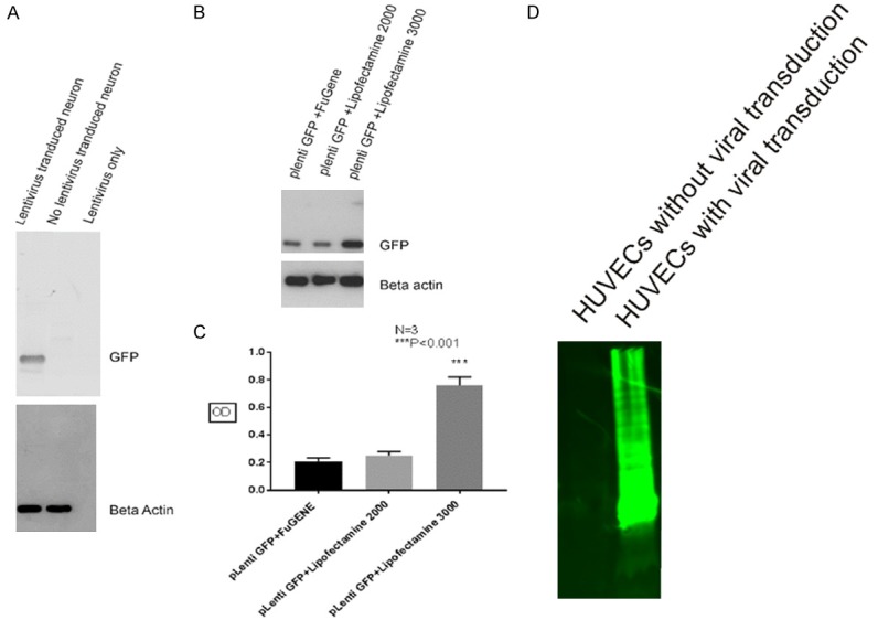
Figure 5.

Western blot showing high level of GFP expression in cortical neuron cell lysates after viral transduction using Lipofectamine 3000 reagent. Using cell lysates from HUVECs after three days of viral transduction with virus generated utilizing Lipofectamine 3000 reagent, western blot showed a strong GFP band migrating from the top of the gel to the bottom after using 10 ug of total protein loading, however, there is absent of GFP detection using cell lysates without viral transduction (D). After SDS-PAGE using 1 ug protein loading, a single protein band migrating around 25 kDa is clearly seen using cell lysates from virus transduced mouse cortical neurons (A, upper panel, lane 1), but the band is absent using cell lysates from mouse cortical neurons without undertaking viral transduction or using supernatant of lentivirus only (A, upper panel, lane 2 and 3), the same membrane was striped and reprobed with anti-beta actin antibody showed equal beta-actin detection (A, lower panel, lane 1 and 2), but there was no beta-actin band detection using supernatant containing lentivirus only (A, lower panel, lane 3). Western blot also showed highest GFP protein expression in mouse cortical neurons using virus generated from HEK293 cells transfected with Lipofectamine 3000 reagent (B, upper panel, lane 3), while less GFP is detected using virus produced with FuGENE 6 or Lipofectamine 2000 transfection reagents (B, upper panel, lane 1 and 2), the same membrane was striped and reprobed with anti-beta actin antibody, which showed equal beta-actin detection (B, lower panel). Similarly, statistics analysis also showed that GFP protein level using lipofectaime 3000 reagent is at least three times higher than using Lipofectamine 2000 or FuGENE 6 transfection reagents (C).
