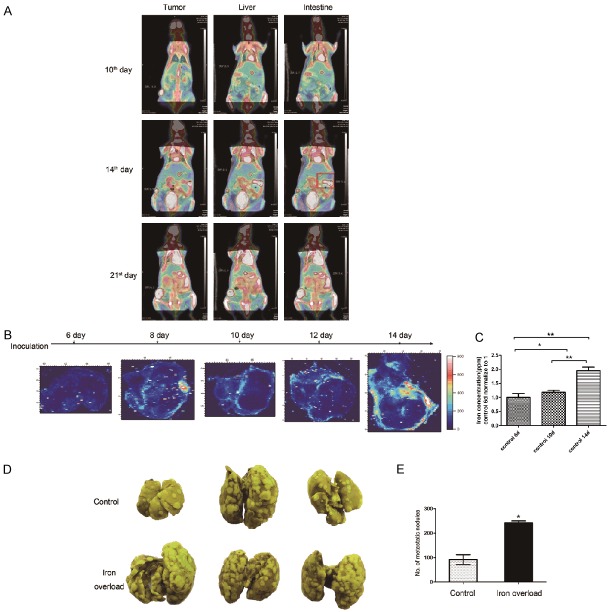
Figure 1.
Iron accumulation was related with tumor metastasis. A. 18F-FDG microPET/CT scan indicated that in subcutaneous 4T1 breast carcinoma model, the first detected foci of metastasis were detected on day 14 after inoculation (intestine, red box). B. SXRF images of iron concentration and distribution at different growth stages after inoculation. Color bar indicated the concentration of iron. C. The iron concentration in tumor tissue was measured by using ICP-MS. D. 21 days after intravenous injection of 105 4T1 cells in mice, lungs were harvested and fixed with Bowie’s solution. Yellow spot indicated the metastatic foci. E. Statistical analysis of the number of metastatic foci observed. *P<0.05, **P<0.01.

