Figure 3.
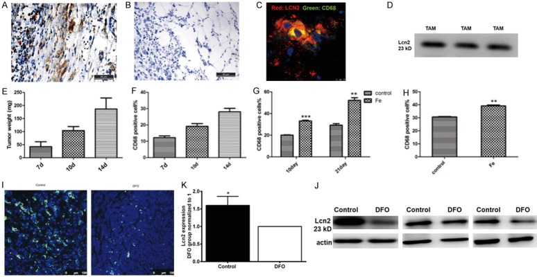
TAMs colocalized with Lcn2 and secreted Lcn2. A. Immunohistochemical staining of Lcn2 (asterisk marks) in the peripheral region of tumor. B. Isotype IgG was used to address the specificity of Lcn2 primary antibody. Scale bar, 50 μm. C. Co-localization of CD68 positive TAMs and Lcn2. Scale bar, 10 μm. red: Lcn2, green: CD68, blue: DAPI. D. Lcn2 was detected in TAMs culture medium by western blot. Three lanes represented three replicates. E. Tumor weights were evaluated on different days after tumor inoculation. F. Percentage of TAMs in tumors tissue at different growth stages. G. Percentage of TAMs after intraperitoneal injection of iron dextran. H. Percentage of TAMs in in situ breast cancer model. I. immunofluorescense staining of CD68 in tumor tissues from control group and DFO group. J, K. The expression of Lcn2 in control and DFO group detected by western blot and the results were repeated three times. n=3. Data was shown as mean ± SD. *P<0.05, **P<0.01 by Student’s t-test.

