
Figure 12.
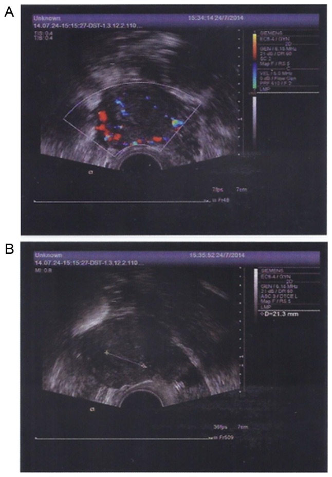
Transvaginal color Doppler flow imaging and transabdominal ultrasound detection of endometrial morphology on July, 24 2014. (A) Abundant blood flow signals in the endometrium were detected. The sizes of the left and right ovary were 3.3×2.0 and 2.7×1.2 cm, respectively. (B) Endometrial thickness was 0.75 cm.