
Figure 2.
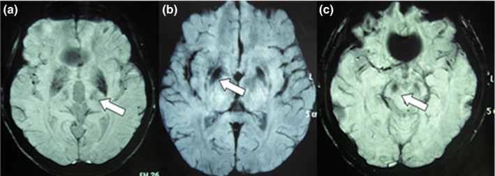
Susceptibility‐weighted imaging results showing low signals of ROIs in the brains of patients with WD. CP values of the TH was lower on the left side than on the right side (a), CP values of the PU were lower on the right side than on the left side (b), CP values of the SN were lower on the right side than on the left side (c). Abbreviations: PU, putamen; TH, thalamus; SN, substantia nigra; ROI, regions of interest; WD, Wilson's disease