Figure 3.
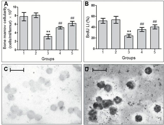
VC-III induced protection against CDDP-induced myelosuppression. Histograms show (A) bone marrow cellularity and (B) BrdU LI (%) in different groups after CDDP administration. Data were represented as mean ± SD, n = 6. **Significantly (P < 0.001) different from Group 1 and ##significantly (P < 0.001) different from Group 3. Representative photomicrographs of cell proliferation assay showing (C) non-proliferating cells and (D) proliferating cells (indicated by BCIP/NBT staining), ×400 magnification, scale bar = 25 μm.

