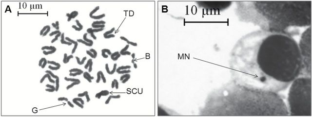
Figure 4.
VC-III mediated prevention of CDDP-induced clastogenesis. (A) Metaphase complements of bone marrow cells showing structural aberrations, ×1000 magnification, scale bar = 10 μm. Arrows indicate break (B), gap (G), sister-chromatid union (SCU) and terminal deletion (TD). (B) Representative photomicrograph of Giemsa-stained bone marrow slide showing MN (indicated by black arrow), ×1000 magnification, scale bar = 10 μm.

