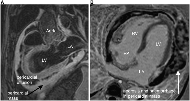
Figure 1.
(A) Three-chamber, T2-weighted image showing large circumferential pericardial mass (black arrow) with pericardial effusion (PE). Left atrium (LA), left ventricle (LV) and aorta are marked. (B) Four-chamber image following late gadolinium enhancement, showing areas of necrosis and haemorrhage within the pericardial mass (marked with white arrow). LV, LA, right ventricle (RV) and right atrium (RA) are marked.

