
Figure 1.
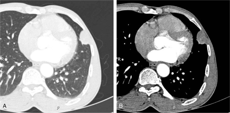
Chest computed tomography-enhanced images of the (A) lung window and (B) mediastinal window. A nodule with a well-circumscribed margin and a heterogeneous enhancement pattern was observed in the left upper lobe in proximity to the left chest wall. Some pulmonary small arteries were observed at the edge of the nodule (B).