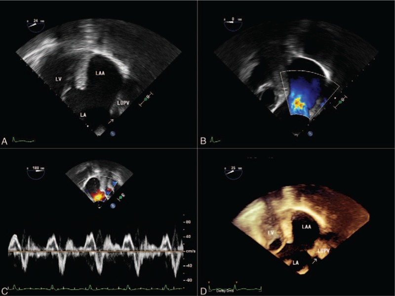
Figure 4.

Transesophageal echocardiograms. The mid-esophageal 2-chamber and LAA view (A) revealing a giant LAA. Color Doppler flow imaging (B) showing communication of flow between LAA and the LA. Pulsed wave Doppler (C) echocardiographic examination of blood flow at the orifice. Three-dimensional view (D) confirming the presence of giant LAA. LA = left atrium, LAA = left atrial appendage, LV = left ventricle, LUPV = left upper pulmonary vein.
