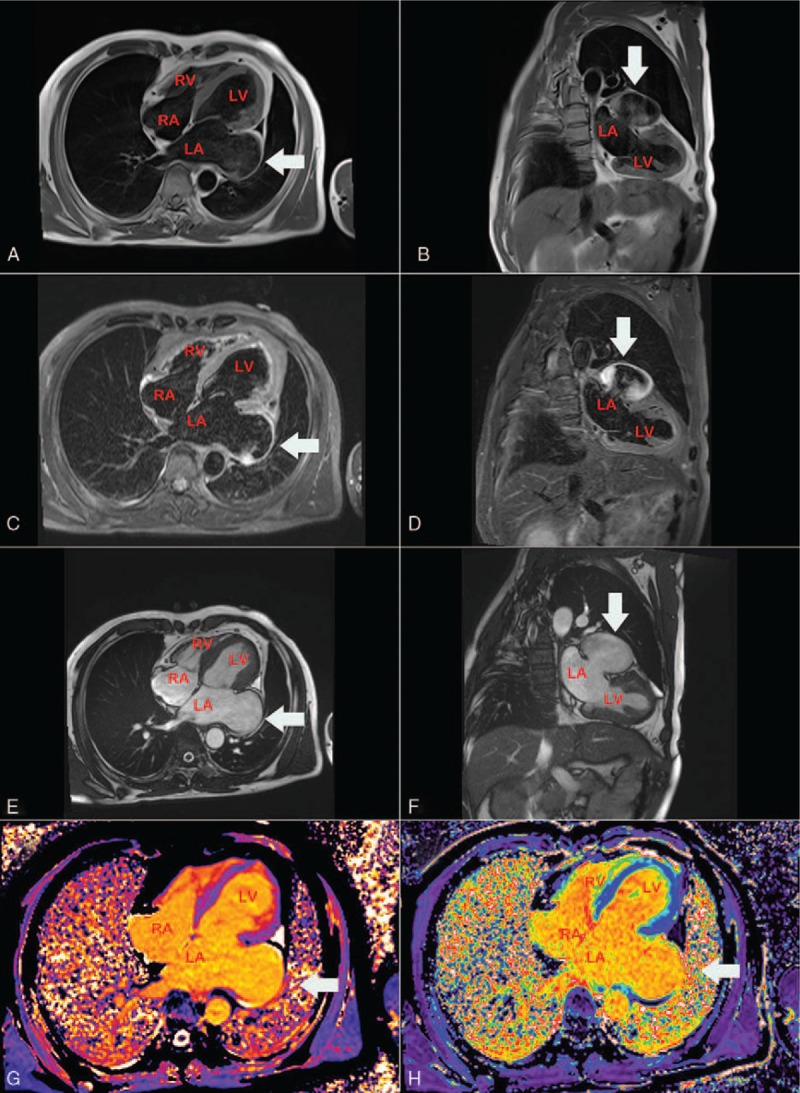
Figure 6.

MRI. Axial spin-echo T1-weighted (E) and Coronal spin-echo T1-weighted image (F) showing the LAA aneurysm with no evidence of thrombus. LA = left atrium, LV = left ventricle, MRI = magnetic resonance imaging, RA = right atrium, RV = right ventricle.
