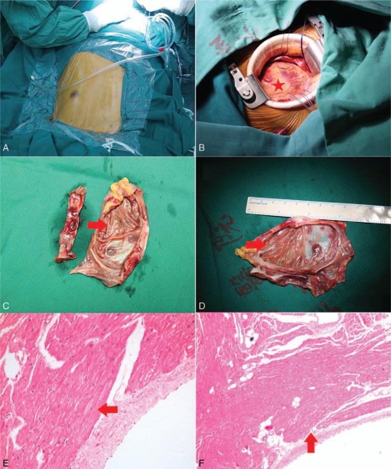
Figure 7.

Intraoperative views (A–D) and histopathological pictures (E, F). The resected aneurysm measured 8.7 × 7.0 cm (C, D) through a limited left thoracotomy (A, B). Histopathological section of the aneurysm showing the myocardial thinning with fatty infiltration.
