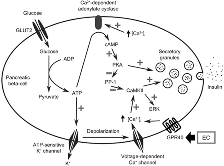
Figure 6.
Schematic diagram of the possible pathways for EC-mediated insulin secretion in pancreatic β-cells. An increased cellular adenosine triphosphate (ATP)/adenosine diphosphate (ADP) ratio primarily induced by oxidative phosphorylation closes ATP-sensitive K+ channels, which subsequently causes membrane depolarization and opening of voltage-dependent Ca2+ channels, leading to increased cytosolic [Ca2+]i. Epicatechin (EC) can activate G-protein coupled receptor (GPR) 40 to further increase cytosolic [Ca2+]i. The increased [Ca2+]i activates Ca2+/calmodulin-dependent protein kinase II (CaMKII), which serves as the triggering signal in glucose-induced insulin secretion and increases extracellular signal-regulated kinase (ERK) phosphorylation. The increased [Ca2+]i also leads to activation of Ca2+-dependent adenylate cyclase, which increases the cyclic adenosine monophosphate (cAMP) level, leading to protein kinase A (PKA) activation. Activated PKA then prevents dephosphorylation of CaMKII by inhibiting protein phosphatase–1 (PP1), which specifically dephosphorylates CaMKII.

