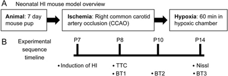
Figure 2.
Schematic of the in vivo neonatal hypoxic-ischemic (HI) mouse model to examine the effects of DCPIB. (A) Overview of the process involved in developing the model, which contained two main parts: ischemia and hypoxia. First, using postnatal day 7 (P7) mice, ischemia was induced by isolation of the right common carotid artery and then subsequent ligation with a bipolar electrocoagulation device (common carotid artery occlusion; CCAO). Following recovery from the surgery, hypoxia was then induced by placing operated pups in a 37°C chamber perfused with a gas mixture of 7.5% oxygen and 92.5% nitrogen for 60 min. (B) Timelines of the experimental sequences for model development as well as the subsequent histological, morphological, and behavioural assessments. The timeline was carried out across three groups: sham, control HI, and DCPIB-treated HI. The “control HI group” represents HI-induced animals administered with vehicle only; “DCPIB-treated HI group” represents HI-induced animals treated with DCPIB. Infarct volume measurement with TTC was carried out 24 h after HI (ie, when pups were P8), whereas measurement with Nissl was 7 days after (ie when pups were P14). Neurobehavioural performance (BT; behavioural test) was assessed at three separate time points; at one, three, and seven days after HI (ie, when pups were P8, P10, and P14, respectively).

