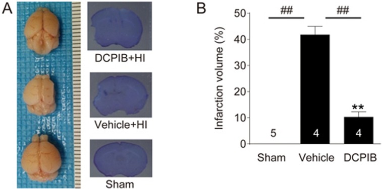
Figure 4.
DCPIB-treated animals showed less morphological and histological damage following HI. (A) Left panel shows representative whole brain images of P14 mouse pups in either the sham group (bottom), control HI group (middle), or DCPIB-treated HI group (top). Note that morphological integrity is severely compromised for the control group brain, when compared to the treatment and sham group brains. Right panel displays the corresponding representative images of Nissl staining. (B) Summary column chart showing that intraperitoneal (ip) treatment of DCPIB (10 mg/kg in 0.2% DMSO) 20 min before induction of HI significantly reduced infarct volume measured seven days following the HI episode (**P<0.01 vs Sham. ##P<0.01 vs vehicle, respectively; n=4/group; One-way ANOVA with Bonferonni multiple comparison test), thus suggesting the lasting neuroprotective effectiveness of DCPIB.

