Figure 2.
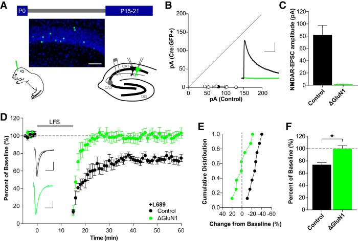
Single-neuron deletion of GluN1 prevents non-ionotropic LTD. A, Schematic of experimental preparation. GluN1fl/fl mice were injected with AAV1-Cre:GFP at P0 for conditional deletion of GluN1. After 15–21 d, dual whole-cell recordings were made from neighboring transduced and control neurons. Representative image of the sparse transduction of CA1 pyramidal cells with AAV1-Cre:GFP counterstained with DAPI. Scale bar, 100 μm. B, C, NMDAR-EPSCs are eliminated by 15–21 d. B, Scatterplot of individual neuron pairs (open circles) and averaged pair ± SEM (solid circle). Sample trace scale bars indicate 100 ms, 40 pA. C, Average NMDAR-EPSC amplitudes for control (82.1 ± 15.7 pA, n = 5) and Cre:GFP+ neurons (1.75 ± 0.53 pA, n = 5; t(4) = 5.021, p = 0.007, paired t test). D–F, Deletion of GluN1 prevents LTD. D, Averaged whole-cell LTD experiments and representative traces (10 ms, 50 pA). E, Cumulative distribution of experiments in D. F, Average percentage depression relative to baseline; control neurons (73.7 ± 3.5%, n = 8), Cre:GFP+ neurons (ΔGluN1: 99.8 ± 5.2%, n = 8; t(14) = 4.194, *p = 0.0009, t test).

