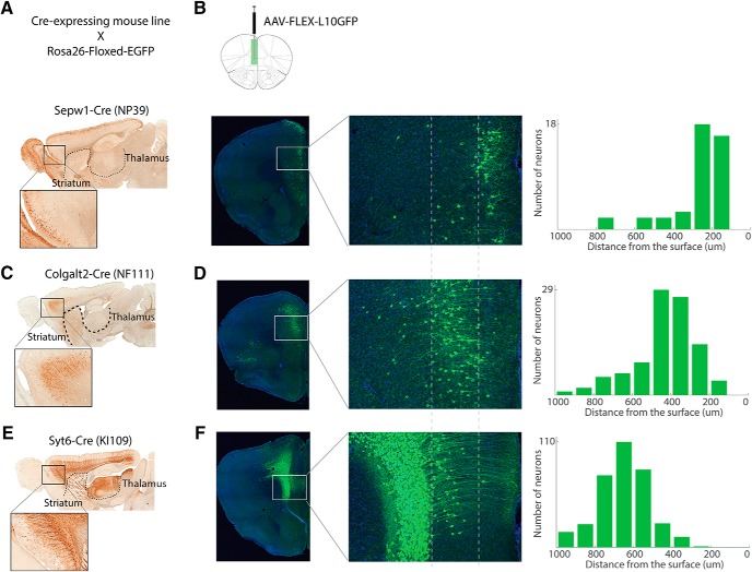
Figure 5.
Cre-expressing transgenic mouse lines that target pyramidal neurons in different cortical layers in the mPFC. A, C, E, Sagittal sections of Cre-expressing mouse lines crossed with the reporter Rosa26-floxed-EGFP reporter line. A, Sepw1-Cre (NP39), (C) Colgalt2-Cre (NF111), and (E) Syt6-Cre (KI109). B, D, F, Left, Location of Cre-positive neurons in the mPFC of each mouse line was visualized by unilateral injection of AAV-FLEX-L10-EGFP virus in Sepw1-Cre (B), Colgalt2-Cre (D), and Syt6-Cre (F). Middle column, Magnified views of images in the left column. Right column, Number of cells along cortical depth in the areas shown in the middle column.

