
Fig. 4.
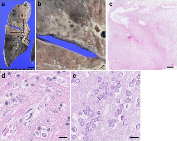
Histopathological findings of right lung. a and b Gross appearance of coronal section of the right lung. Grayish lesion with clear boundary was observed in the upper lobe of the right lung. b shows the magnified image of square area in (a). Scale bar: 2 cm. c Very low-power field of the right lung. Necrotic lesion with clear boundary was observed. Scale bar: 2 mm. d and e High-power field of necrotic lesion in the right lung. Amoebic cysts were observed in a part of nectoric lesion (d). These cysts showed faint positive in Periodic acid-Schiff stain (e). Scale bar: 20 μm