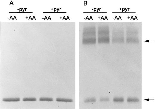
Figure 6.
Immunoblot of the AOX protein present in mitochondria isolated from transgenic (B9) tobacco suspension cells d 3 after subculture. In some cases, cells were pretreated for 1 h with 10 μm AA prior to the mitochondrial isolation. Media used in the mitochondrial isolation were supplemented with 5 mm pyruvate (+pyr) or were not supplemented (−pyr). Mitochondrial proteins (100 μg) were resolved by either reducing (A) or non-reducing (B) SDS-PAGE prior to the immunoblot analysis shown. Arrows indicate the position of the oxidized (upper arrow) and reduced (lower arrow) forms of the AOX protein. Representative results are shown.

