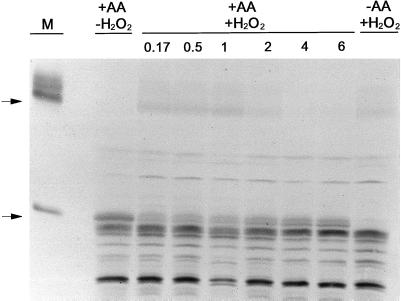
Figure 9.
Immunoblot of the AOX protein present in a cellular protein extract from wild-type tobacco suspension cells. Cells d 3 after subculture were either left untreated (−AA) or were treated for 24 h with 10 μm AA (+AA) to induce large amounts of AOX protein in the cells. Then, the cells in culture were treated with 5 mm H2O2 (+H2O2). At different times following the H2O2 addition (ranging from 0.17 to 6 h), a cellular protein extract was obtained from an aliquot of the cell culture, and 40 μL of the extract was subsequently analyzed by non-reducing SDS-PAGE and immunoblot analysis. The lane marked “+AA, −H2O2” is a protein extract from cells just prior to the H2O2 addition. The lane marked “−AA, + H2O2” is an extract from cells not given the pretreatment with AA prior to the 10-min incubation with H2O2. The lane marked “M” is a protein sample from isolated mitochondria to clearly show the position of the oxidized and reduced forms of the AOX protein. The arrows at left indicate the position of the oxidized (upper arrow) and reduced (lower arrow) forms of the AOX protein.

