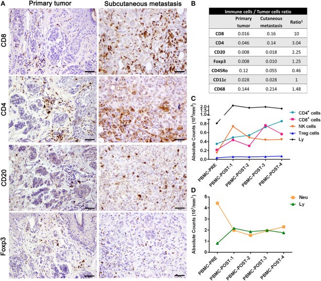
Figure 2.
Immune profiling of patient #006. (A) Representative pictures of the primary tumor and subcutaneous metastasis biopsies infiltrated by CD8+, CD4+, CD20+, and Foxp3 lymphocytes determined by IHC (DAB, brown). (B) After counting total biopsy area, immune-to-tumor cell ratios were determined for each tumor biopsy as well as the relative ratio between primary tumor and SC metastasis (1). (C) Absolute counts of total lymphocytes, T CD4+, T CD8+, Treg, and NK cells from PBMC were determined throughout CASVAC-0401 treatment and follow-up by flow cytometry; also, absolute counts of lymphocytes (Ly) and neutrophils (Neu) are shown (D). Scale bars = 50 µm.

