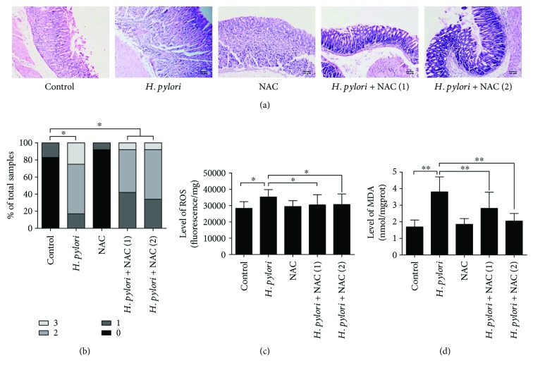
Figure 3.
H. pylori infection induces gastritis and oxidative damage in the gastric tissue of Balb/c mice. Gastric tissue sections from H. pylori-infected mice were taken after 24 weeks of infection. (a) Pathological changes were evaluated by HE staining. (b) Gastritis histopathology was graded according to the updated Sydney System. (c) Gastric ROS levels were determined by measuring DCF fluorescence. (d) MDA levels in the gastric mucosa were detected. Scale bar = 50 μm; ∗p < 0.05 and ∗∗p < 0.01.

