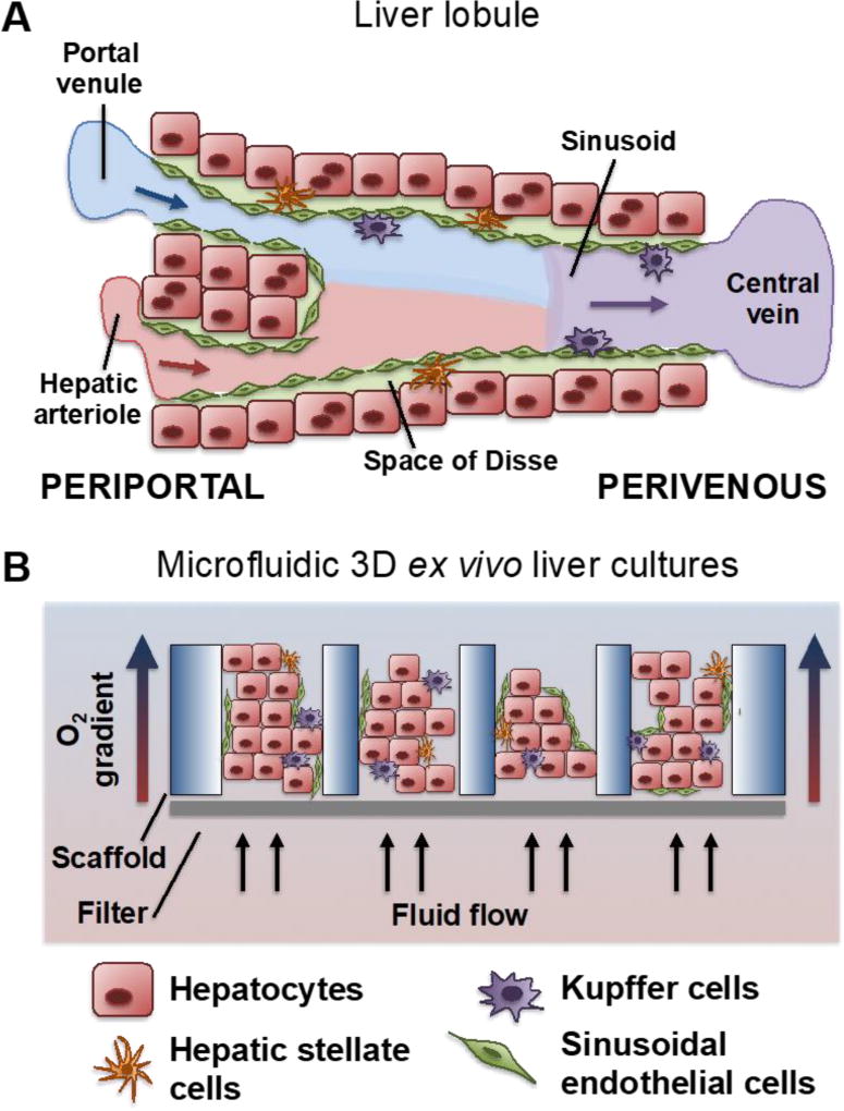
Figure 1.
(A) The architecture of the hepatic sinusoid. Blood enters the portal triad region of the liver through a hepatic arteriole and portal venule and traverses the hepatic sinusoid to the central vein, whereby it is drained into the larger hepatic central veins. The sinusoidal endothelial cells mediate blood flow and are fenestrated to allow rapid diffusion of nutrients, signaling factors, and drug compounds. The hepatocytes compose the parenchyma of the liver and sit deep to the endothelium. The hepatic stellate cells reside in the Space of Disse, a zone between the endothelium and hepatocytes. Finally, the Kupffer cells line the inside of the sinusoid and mediate antigen sensing and intercellular communication. (B) A schematic of the three dimensional liver tissues as presented within the LiverChip, that are subject to fluid flow in micro-scale pores of a scaffold, which facilitates shear stresses and establishment of a physiological oxygen gradient.

