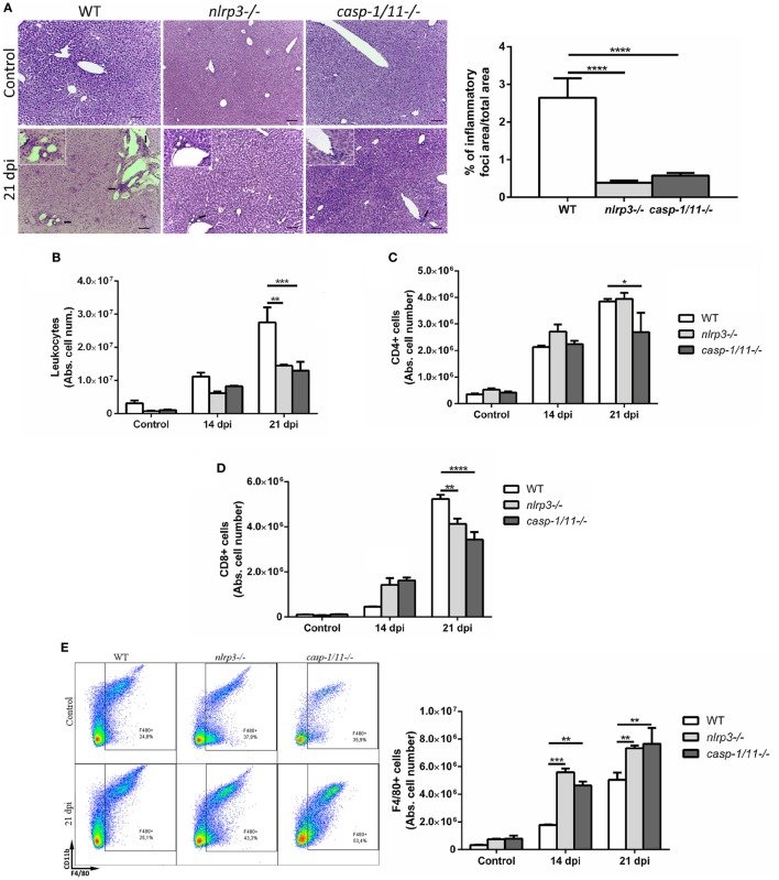
Figure 2.
Infected caspase-1/11-deficient mice exhibit a marked reduction of hepatic CD8+ T cell number associated with increased number of macrophages. (A) Representative liver sections from uninfected control and Trypanosoma cruzi infected wild type (WT) and KO mice at 21 days post infection were stained with hematoxylin and eosin. Micrographs are shown at 100× and bar scales depict 50 µm. Insets are magnifications of inflammatory infiltrates. Bars depict percentages of inflammatory foci. (B) Absolute number of hepatic leukocytes from WT, nlrp3−/−, and caspase-1/11−/− mice determined by flow cytometry. (C) Absolute number of purified hepatic CD4+ and (D) CD8+ T cells. (E) Representative dot plots and absolute cell number of F4/80+CD11b+and F4/80+CD11b− leukocytes. Gate strategy is depicted in Figure S1 in Supplementary Material. Data are shown as mean ± SEM from one of three representative experiments; n = 3–6 mice per group. Statistical significance was evaluated by two-way ANOVA followed by Bonferroni post hoc test. *p < 0.05; **p < 0.01; ***p < 0.001.

