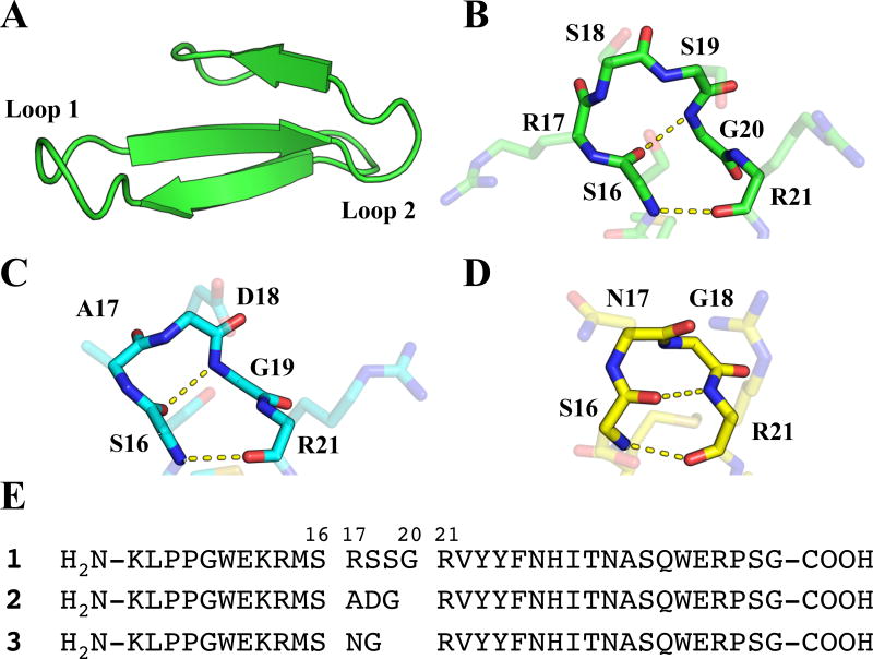
Figure 1.
(A) Structure of the isolated Pin1 WW domain, derived from PDB entry 4GWT. (B) View of Loop I within the Pin1 WW domain with backbone atoms (N, Cα, C, O) highlighted. Intramolecular hydrogen bonds correspond to a 4:6 β-hairpin classification. (C) View of the 3:5 β-hairpin from WW domain variant 2 (PDB: 2F21), with intramolecular hydrogen bonds shown. (D) View of the 2:2 β-hairpin from WW domain variant 3 (PDB: 1ZCN), with intramolecular hydrogen bonds shown. (E) Sequences of WW domain peptides 1, 2, and 3.

