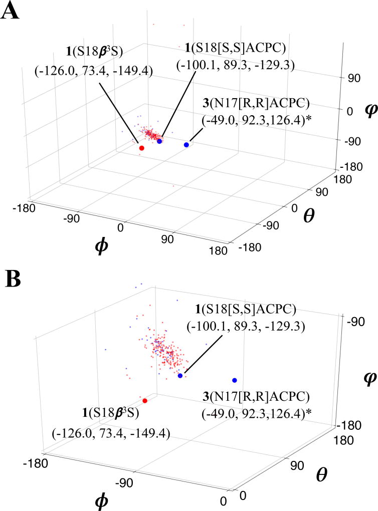
Figure 5.
Global (A) and zoomed-in (B) views of β3-residue (red; 185 shown) and ACPC (blue; 46 shown) backbone geometry in φ/θ/ψ space, from previously-reported structures (see Supporting Information for relevant PDB accession codes) and three WW domain x-ray structures reported here. Backbone angles corresponding to the new WW domain structures are shown as large red or blue circles. *Note that in both plots, signs of backbone dihedral angles for [R,R]ACPC in structure of 3(N17[R,R]ACPC) have been reversed to facilitate comparison with angles derived from “right-handed” residues.

